Assessment and feedback
Feedback for Part 1 - Core Concepts
End of Part 1 - Core Concepts

Paleoradiography of human dry bones
The radiographic survey of 92 medieval and post medieval skeletons from St Albans, United Kingdom
A research project funded by the College of Radiographers Industry Partnership Scheme.
An account of the paleoradiographic survey of archaeological bones from St Albans, United Kingdom.
James Elliott - November 2021
Radiographic technique for archaeological human dry bones
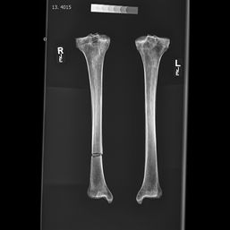
This project investigated the radiographic technique and workflow used to survey 92 medieval and post- medieval skeletons from St Albans, United Kingdom. As a dual qualified archaeologist and diagnostic radiographer, I was interested to find out if there were specific recommendations for imaging human dry bones for archaeological analysis. At present there seems to be scarce literature on the topic, so I teamed up with Adelina Teoaca, an osteoarchaeologist from Canterbury Archaeological Trust, to find out the 'best' technique. We carried out a comprehensive radiographic survey of long bones to identify biological stress (as Harris lines) and develop a process for future investigators.
The findings have been published as a journal article within Radiography and as a conference presentation for the British Association for Biological Anthropology and Osteoarchaeology. Both the article and a video of the conference presentation are available below.
KEY STATISTICS
-
502 x-rays taken of 426 long bones.
-
Over 5 days April-May 2021
-
Team: Diagnostic radiographer and osteoarchaeologist
-
Sample: Medieval 1066-1485 and Post Medieval from an excavation of Monk's Graveyard, St Albans, United Kingdom
Main findings:
These recommendations are based upon our experiences during the survey and may be of benefit to other investigators.
1. Use of a digital radiography system. This allowed the rapid acquisition of imaging during the survey and provided digital files (DICOM) for easy storage, sharing and analysis. Although not listed within the recommendations of the published paper, the speed of imaging is noted within the discussion.
2. At a minimum, the team should comprise of a radiographer and osteoarchaeologist. Cabinet x-ray units, such as the Faxitron from Wessex Archaeology, do not necessarily need a radiographer to perform the imaging. However, our imaging made use of a clinical x-ray room that provided excellent positioning of specimens and customisation of radiographic technique. In any case, the input of a radiographer should be considered. A dedicated photographer may also be of benefit.
3. Diligent records and accurate annotation of radiographs. This acted as a failsafe for future analysis of the imaging, without which the validity and reliability of the imaging could be questioned.
The back story
In April 2020 I applied for the College of Radiographers Industry Partnership Scheme (CoRIPS) for £5000 to pay for research time and some equipment. Although not directly related to archaeology, CoRIPS supports investigations related to forensic radiography. Archaeological remains have little direct relevance to forensic investigation, as they lack the legal importance associated with questions of the law, however they provide an opportunity to test methodologies and process.
By pure coincidence, my colleague Adelina Teoaca required my assistance with her Masters thesis to investigate biological stress in a medieval and post-medieval population of remains. We collaborated together to image all the long bones of 92 individuals from an excavation in St Albans. With CoRIPS paying for my time, I used the radiography equipment at Canterbury Christ Church University to locate Harris lines as evidence of nutritional or pathological stress. The imaging took place over five days in April-May 202.
The future?
The results of the Harris line investigation will be published at a later date, Adelina has only recently graduated! We hope to data-mine the x-rays to learn more about the deceased: photodensitometry (density estimation), radiogrammetry (cortical thickness as an indicator of bone loss) and radiometrics (using radiographs for direct measurement). I also wish to use the images within teaching.
The published reflective account is shown below, it has not yet been assigned a volume. It is Open Access, free to distribute. The direct link is https://doi.org/10.1016/j.radi.2021.10.011
Conference presentation
In September 2021 the British Association for Biological Anthropology and Osteoarchaeology held their annual conference online. The video below provides the entire presentation, including additional photographs and videos of the x-ray room.
More YouTube videos:
Subscribe to YouTube channel:
A supplementary document to the published article is shown below. The document provides additional photographs of the x-ray room, with examples of the radiographic technique deployed during the survey. Again, this is open acces and can be downloaded here. Feel free to distribute with due accreditation.


